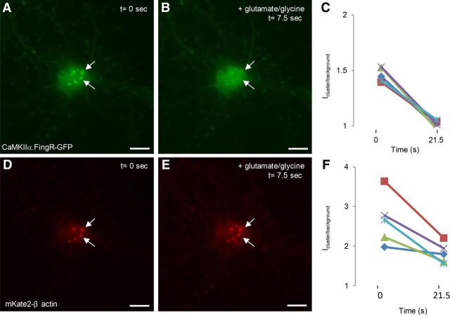
Figure 10.
CaMKIIα clusters dissociate from F-actin clusters upon stimulation. A, Clusters labeled with CaMKIIα.FingR-GFP (green) in cortical neurons. B, Addition of 50 μm glutamate and 5 μm glycine for 7.5 s results in dispersal of the clusters. C, Icluster/background for CaMKIIα.FingR-GFP shows that the clusters have completely dispersed within 21.5 s. D, Clusters of β actin fused to mKate2 in the same cell as in A. E, Addition of 50 μm glutamate and 5 μm glycine for 7.5 s does not disperse the clusters. F, Time course of Icluster/background for mKate2-β actin during exposure to 50 μm glutamate and 5 μm glycine. Scale bars, 10 μm.

