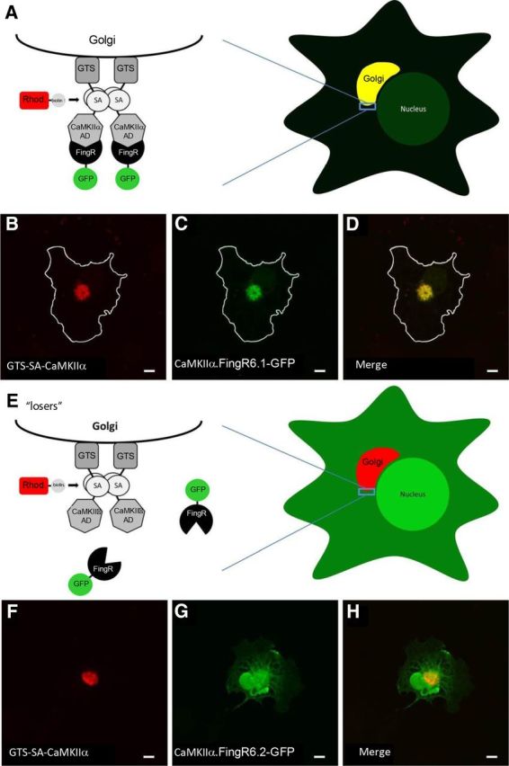
Figure 2.

Intracellular Golgi localization assay for FingR/target binding. A, Schematic shows CaMKIIα target targeted to the surface of the Golgi through a Golgi targeting signal (GTS) following expression in COS-7 cells. Coexpressed FingRs that bind to the CaMKIIα target are colocalized with the target at the Golgi. B, C, COS-7 cell coexpressing a Golgi-targeted CaMKIIα association domain (B, GTS-SA-CaMKIIα, red) and a FingR that binds to CaMKIIα (C, CaMKIIα.FingR6.1-GFP, green). D, Both GTS-SA-CaMKIIα and CaMKIIα.FingR6.1-GFP colocalize at the Golgi. E, Schematic of a FingR that does not bind to target following expression in COS-7 cells. F, GTS-SA-CaMKIIα (red) targeted to the Golgi. G, CaMKIIα.FingR6.2-GFP (green) is localized nonspecifically and does not colocalize with GTS-SA-CaMKIIα. H, Merged image shows that CaMKIIα.FingR6.2-GFP and GTS-SA-CaMKIIα do not overlap, indicating that the FingR does not bind at high affinity to the target. Scale bars, 5 μm.
