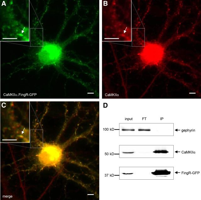
Figure 3.
CaMKIIα.FingR-GFP colocalizes with endogenous CaMKIIα in neurons. A, B, CaMKIIα.FingR-GFP (A, green) expressed in a dissociated cortical neuron colocalizes similarly to CaMKIIα (B, red). C, Yellow color of merged image of CaMKIIα and CaMKIIα.FingR-GFP indicates colocalization of the two proteins. D, Immunoprecipitation of CaMKIIα.FingR-GFP expressed in cortical neurons in dissociated culture results in coprecipitation of virtually 100% of endogenous CaMKIIα, indicated by its absence in flow through (FT). In contrast, endogenous gephyrin is not coprecipitated. Scale bars, 5 μm.

