Figure 7.
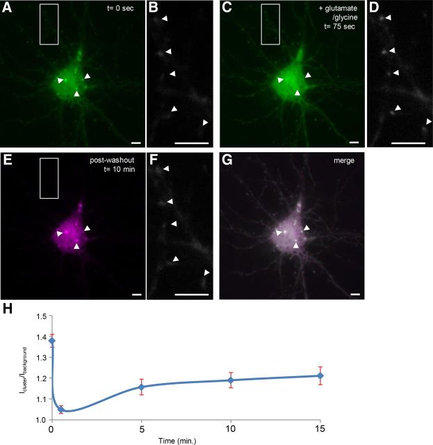
Following dispersal, clusters labeled with CaMKIIα.FingR-GFP re-form in their original positions and shapes. A, Clusters labeled with CaMKIIα.FingR-GFP in a cortical neuron in culture. B, Dendrite from the same neuron as in A shows faint, diffuse staining by CaMKIIα.FingR-GFP. C, Clusters in A dispersed following addition of 50 μm glutamate and 5 μm glycine. D, CaMKIIα.FingR-GFP labeled tight clusters in same dendrite as in B following addition of 50 μm glutamate and 5 μm glycine. E, Following washout of glutamate and glycine and incubation for 10 min, the clusters re-formed in their original positions and shapes. F, Same dendrite as in B and E showed diffuse labeling by CaMKIIα.FingR-GFP following washout of glutamate and glycine. G, Superimposition of the images of clusters before and after dispersal and re-formation confirms that they re-formed in the same locations and configurations as the original clusters. H, Time course of Icluster/background for CaMKIIα.FingR-GFP in the presence (t = 0 to t = 75 s) and absence (t > 75 s) of glutamate and glycine. Error bars represent SEM. Scale bars, 10 μm.

