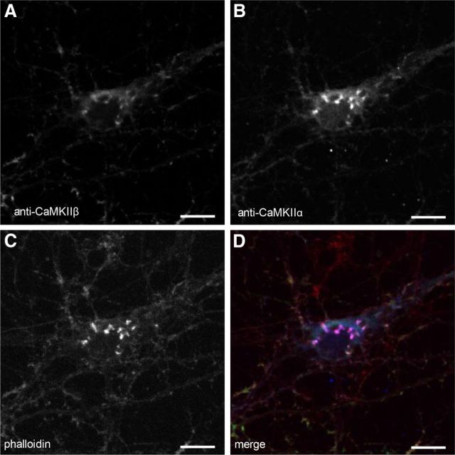
Figure 8.
CaMKIIα and CaMKIIβ colocalize with F-actin in clusters within the cell body. A, Cluster in the cell body of a cortical neuron in culture stained with anti-CaMKIIβ antibody. B, C, Same cell as in A colabeled with anti-CaMKIIα (B) or with phalloidin (C). D, Merge confirms that expression patterns of CaMKIIα (blue), CaMKIIβ, (green), and actin (red) overlap. Scale bars, 10 μm.

