
Fig. 1.
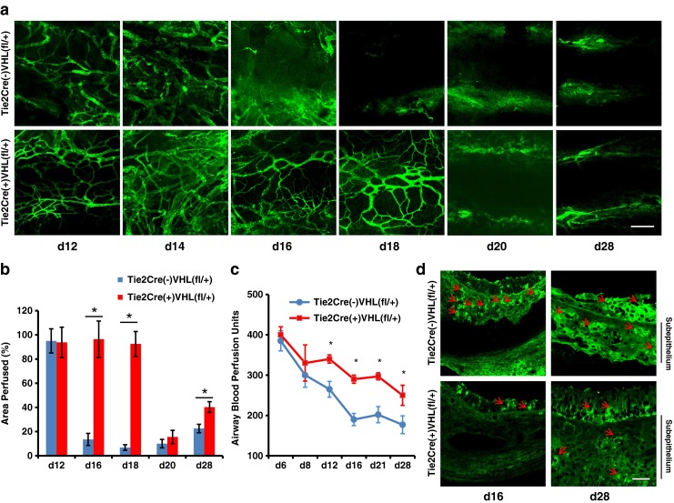
Recipient Tie2+ cell VHL haplodeficiency prolongs airway microvascular perfusion and diminishes tissue hypoxia. a Confocal microscopic FITC-lectin perfusion images showing microvascular perfusion of tracheas transplanted into control recipients with normal VHL expression compared with recipients with VHL haplodeficiency in Tie2+ cells. b Quantification of perfused airway microvasculature following transplantation (n = 4–6). c Airway blood perfusion measured by laser Doppler flowmetry in tracheas transplanted into recipients with or without VHL haplodeficiency (n = 4–6 per time point). d Pimonidazole staining (green, red arrows) in transplanted airways in both day 16 and day 28 showing decreased hypoxia in grafts transplanted into VHL-haplodeficient recipients. Scale bars, 100 μm (a and d). Data are shown as means ± SEM. *P < 0.05 at individual time points, Student’s t test