
Fig. 4.
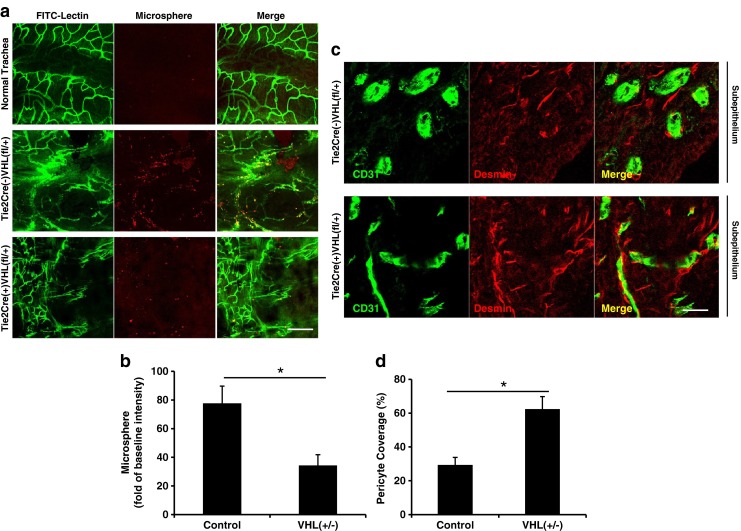
Improved vessel function and maturation in airways transplanted into recipients with Tie2+ cell VHL haplodeficiency. a Representative confocal images showing extravasated 50 nm microsphere (red) and microvascular perfusion (green) in normal airways and airways transplanted into control or VHL-haplodeficient recipients at day 28. b Quantification of microsphere extravasation (n = 4–6). c CD31 (marker for ECs) and desmin (marker for pericytes) staining showed more complete pericyte-covered vessels in airways transplanted into VHL-haplodeficient recipients at day 28. d Quantification of pericyte coverage in the microvasculature of transplanted airways (n = 3–5). Scale bars, 100 μm (a), 20 μM (c). Data are shown as means ± SEM, *P < 0.05, Student’s t test