Fig. 5.
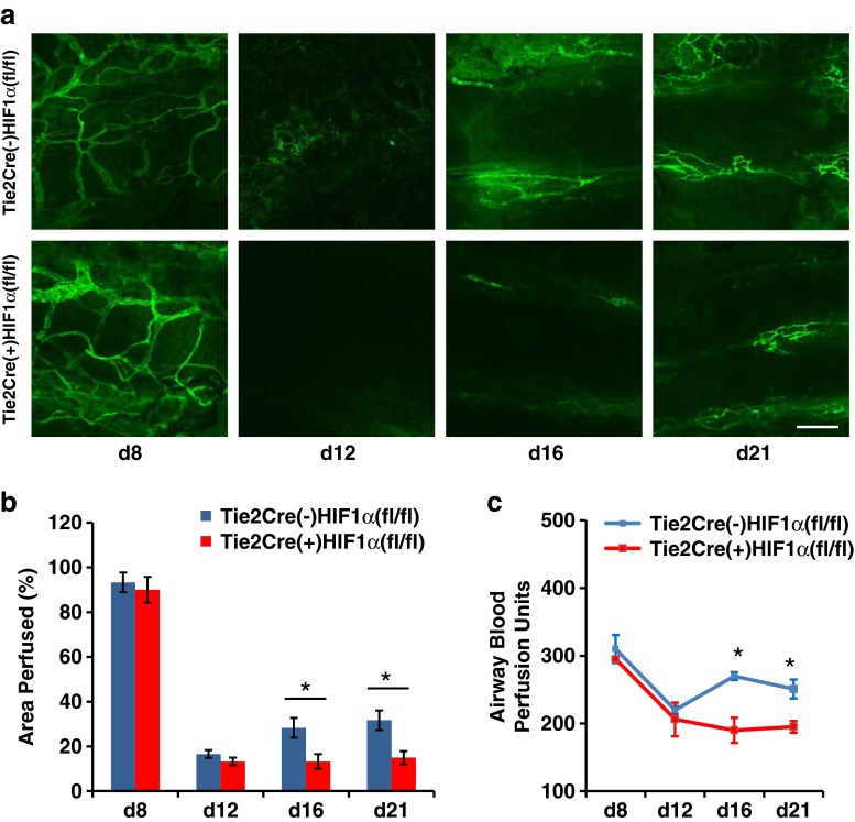
Diminished microvascular perfusion of airways transplanted into recipient with HIF-1α knockout in Tie2 lineage cells. a Representative FITC-lectin microvascular perfusion images at various time points posttransplantation. b Quantification of relative microvascular perfusion (n = 4–6). c Laser Doppler flowmetry analysis showing relative perfusion of tracheal transplants (n = 4–6 per time point). Scale bar, 100 μm (a). Data are shown as means ± SEM. *P < 0.05, Student’s t test

