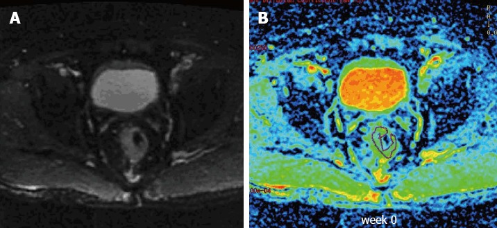
Figure 1.

Sample T2-weighted and diffusion-weighted images from a patient with locally advanced rectal cancer prior to treatment. A: T2-weighted transaxial image through the pelvis prior to treatment; B: Corresponding apparent diffusion coefficient map of the patient obtained from the diffusion-weighted images. A region of interest was drawn around the tumor.
