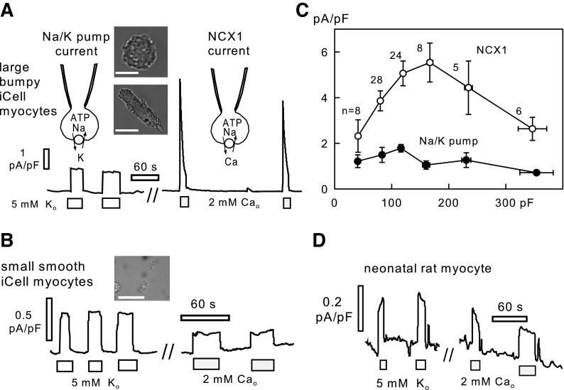
Fig. 2.
Representative Na/K pump and Na/Ca exchange current recordings from iCell Cardiomyocytes and rat neonatal cardiomyocytes at 37°C. A: recordings from a large iCell Cardiomyocyte with well-developed myofilaments. The 2 micrographs illustrate the range of morphologies of large iCell Cardiomyocytes. Scale bars = 50 μm. B: recordings from a round iCell Cardiomyocyte with more fibroblast-like morphology in bright field microscopy. Scale bar = 50 μm. C: average Na/K pump and Na/Ca exchange (NCX1) current densities from 79 recordings, plotted in dependence on the iCell Cardiomyocyte capacitance (Cm). Data points were binned by Cm magnitude as follows: 35–50 pF, 50–100 pF, 100–150 pF, 150–200 pF, 200–250 pF, and 250–400 pF. D: recording of Na/K pump and NCX1 currents in a rat neonatal cardiomyocyte under identical conditions.

