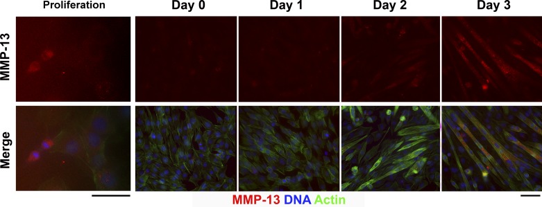
Fig. 3.
Immunocytochemistry for MMP-13 in proliferating and differentiating C2C12 cells. Top: MMP-13 staining alone (red). Bottom: merged image with phalloidin-Alexa 488 (staining actin, green) and 4′,6-diaminido-2-phenylindole (DAPI, blue) to reveal nuclei. Note increased MMP-13 in the daughter nuclei upon cell division (Proliferation, left). MMP-13 is not evident until day 2 of differentiation, after which myotubes exhibit positive cytoplasmic staining. Scale bars, 50 μm.

