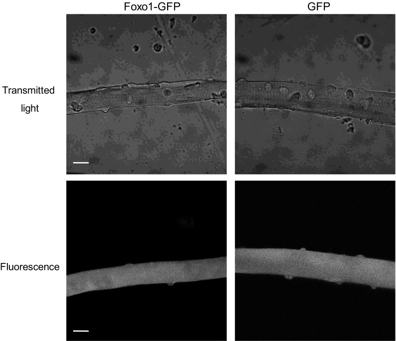
Fig. 1.
Fibers expressing Forkhead box O 1 (Foxo1)-green fluorescent protein (GFP) appear normal. Transmitted light images (top) and GFP fluorescence images (bottom) of Foxo1-GFP (left) or GFP (right) expressing flexor digitorum brevis (FDB) fibers. Signs of health are visible: striations and nuclei are predominantly peripheral, and fibers are smooth and straight. Scale bars = 20 μm.

