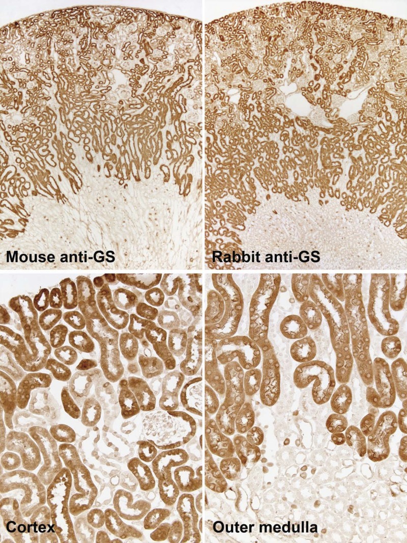
Fig. 2.
GS immunolabel in the mouse kidney. Top: low-power micrographs of normal mouse kidneys demonstrating GS immunolabel using two different primary antibodies against GS, mouse monoclonal antibody (left) and rabbit polyclonal antibody (right). Results were virtually identical with the two antibodies, with expression evident in the majority of epithelial cells in the cortex and OMo and in a small subpopulation of cells in the OMi. Bottom: higher-power micrographs of the cortex and outer medulla. In the cortex, strong immunolabel was present in the majority of tubules, which are composed of cells that have an apical brush border and are identifiable as proximal tubules. In a subpopulation of tubules, there was heterogeneous GS immunolabel, with intense expression in a subpopulation of cells. In the OMo, strong immunolabel in proximal straight tubules (PSTs) was evident. In addition, throughout the outer medulla, a subpopulation of collecting duct cells expressed strong GS immunolabel.

