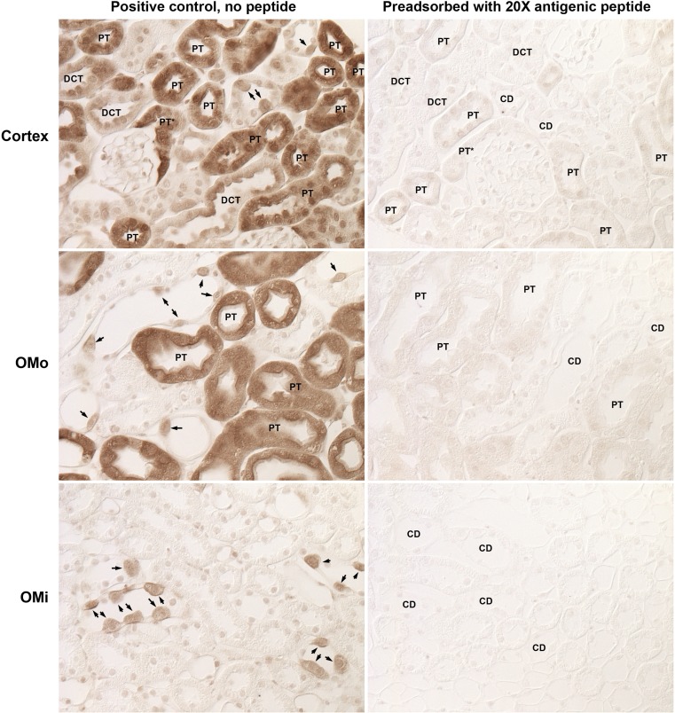
Fig. 4.
Effect of immunizing antigen preincubation with antibody on immunolabel. Mouse kidney sections underwent immunohistochemistry using either rabbit anti-GS primary antibody alone or primary antibody preadsorbed with a 20× excess of the antigenic peptide. Left: representative images from the cortex, OMo, and OMi from kidneys labeled with primary antibody alone. Right: representative images of the corresponding regions from kidneys labeled with primary antibody preadsorbed with excess immunizing peptide. Immunolabeling was virtually eliminated by peptide blocking. Kidneys were photographed using DIC optics to avoid the need for counterstaining to identify cellular profiles. PT, proximal tubule segments; DCT, distal convoluted tubule segments; CD, collecting duct segments. Arrows identify intercalated cells.

