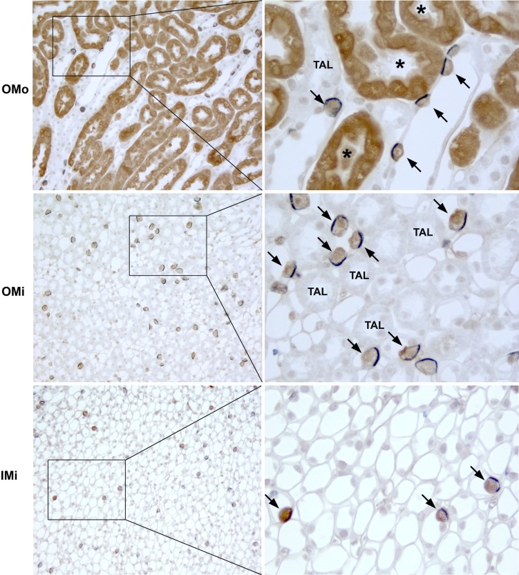
Fig. 5.
GS and anion exchanger 1 (AE1) double label in the medulla. Left: low-power micrographs of the OMo, OMi, and initial inner medulla (IMi) of mouse kidneys labeled for GS (brown) and AE1 (blue) using immunohistochemistry. Right: selected areas (boxes) at higher magnification. There was a 1:1 correlation between GS immunolabel and basolateral AE1 immunolabel expression (arrows) in the outer stripe of the outer medullary collecting duct, inner stripe of the outer medullary collecting duct, and inner stripe of the inner medullary collecting duct, indicating that GS is expressed in type A intercalated cells throughout the medullary collecting duct. GS expression was also evident in PST segments (*). No significant GS immunolabel was evident in other cells in the collecting duct or in the thick ascending limbs TAL.

