
Fig. 3.
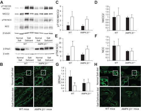
Expression, localization, and phosphorylation of distal Na+ transporters in AMPK-β1−/− mice. AMPK β1−/− mice and littermate control mice were fed the ND or SD for 7 days. A: Western blot analysis of kidney protein lysates. B: immunofluorescence microscopy of kidney sections showing increased subapical expression of Na+-K+-2Cl− cotransporter 2 (NKCC2; SPC-401D antibody) in AMPK-β1−/− mice on the ND compared with WT control mice. C–G: densitometry of Western blots from mice on the ND (solid bars) and SD (open bars). n = 5–10. C: pThr105 (pT105) of NKCC2. D: NKCC2. E: pThr58 (pT58) of the Na+-Cl−1 cotransporter (NCC). F: NCC. G: β-subunit of the epithelial Na+ channel (ENaC). *P < 0.05 and **P < 0.001 for mice on the SD compared with mice from the same strain on the SD; #P < 0.05 for AMPK-β1−/− mice relative to WT mice on the same diet. H: immunofluorescence microscopy of kidney sections showing increased subapical expression of β-ENaC in AMPK-β1−/− mice on the ND compared with WT control mice.