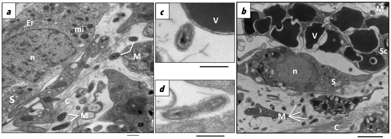
Figure 3. Transmission electron microscopy of thin sections of C. crambe tissues.
Microorganisms in the mesohyl of C. crambe embedded in the collagen matrix of the sponge, (a). Proximity between microorganisms and spherulous cells, (b–c). Bacteria in division, (d). C: collagen; Er: endoplasmic reticulum; mi: mitochondrion; M: Microorganisms; n: nucleus; S:sponge cell; Sc: Spherulous cell whose vesicles (V) contain an electron dense material.

