Fig. 2.
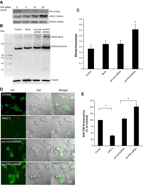
PIN decreases the catalytically active form of nNOS in NG108 neurons. A: knockdown of PIN in NG108 cell line using siRNA. B: estimation of monomers and dimers of nNOS in NG108 cells transfected with Lipofectamine alone (Mock) or plasmid constructs: psi-PIN-shRNA and psi-null-shRNA. After 48 h, dimeric and monomeric nNOS were separated by LT-PAGE in a cold room and visualized by immunoblotting using an anti-nNOS antibody. C: densitometry analyses of monomer and dimer protein levels represented as a ratio of dimer to monomer. *P < 0.05 vs. psi-null-shRNA group. D: representative pictures of intracellular nitric oxide (NO) as measured by 4-amino-5-methylamino-2′,7′-difluorofluorescein diacetate (DAF-FM) in NG108 cells of 4 experimental groups (control, ANG II 100 μM, psi-null-shRNA, and psi-PIN-shRNA). E: cumulative data represented as percent change in NO compared with control without any treatment (n = 8–10 cells from 3 coverslips in each group).

