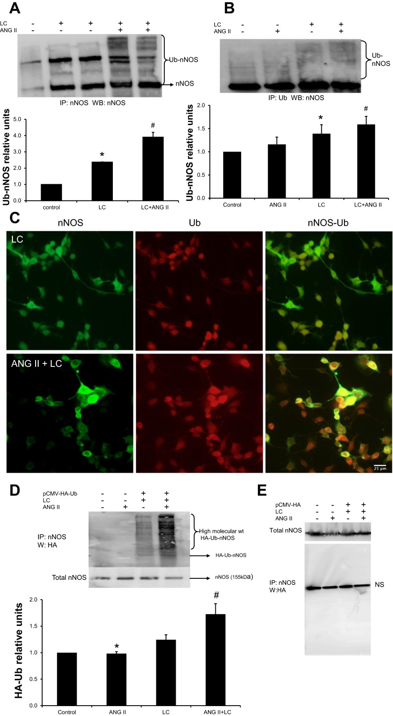
Fig. 6.
ANG II increases the accumulation of higher-molecular-weight forms of immunodetectable nNOS in NG108 cells. A: immunoprecipitation of nNOS from cells treated with ANG II (100 μM), lactacystin (LC, 10 μM), or both was carried out and immunoblotted with anti-nNOS. Top: a representative Western blot; bottom: densitometric analyses of Ub-nNOS bands. Values are means ± SE from 4 independent experiments. *P < 0.05 vs. control, #P < 0.05 vs. LC. B: IP of nNOS from cells treated with ANG II, LC, or both with anti-Ub antibody followed by Western blot with anti-nNOS. Top: a representative Western blot; bottom: densitometry analyses of Ub-nNOS bands. Values are plotted as means ± SE from 4 independent experiments. *P < 0.05 vs. control, #P < 0.05 vs. LC. C: immunocytochemical localization of nNOS (green) and Ub (red) in LC (control) and ANG II along with LC-treated NG108 cells. IP of nNOS-Ub with an anti-nNOS antibody from NG108 cell lysates transfected with pCMV-HA-Ub (D) or control vector (E) and treated with ANG II followed by a Western blot with anti-HA. Top: a representative Western blot; bottom: densitometry analyses of Ub-nNOS bands. A representative experiment distinctive of 4 independent experiments is shown. Values were means ± SE from 4 independent experiments. *P < 0.05 vs. control, #P < 0.05 vs. LC.

