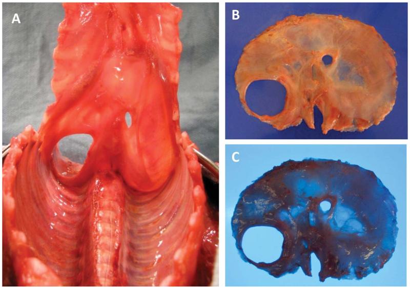
Figure 5.
Photos from a newborn during autopsy with left congenital diaphragmatic defect with thoracic-peritoneal communication exposed using the modified dissection method recommended in the text, which, by reflecting the sternum from the rostral end, does not disrupt the anterior diaphragm. The entire diaphragm should be visualized, examined, and photographed in situ (A, view from the thoracic side). It should next be carefully dissected off the chest wall, removed, and photographed with overhead light (B) and with transillumination (C). A color version of this figure is available online.

