
FIG. 2.
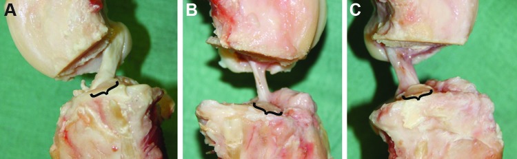
(A) Medial side view of a normal ACL. (B) A healing ACL after suture repair only. (C) A healing ACL after suture repair and SIS. Brackets indicate area of normal insertion site. It can be seen that the treated ACLs do not cover the whole area of normal insertion site. SIS, small intestinal submucosa. Color images available online at www.liebertpub.com/tea