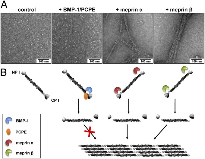
Fig. 2.
De novo fibrillogenesis of type I collagen after cleavage by meprin α or meprin β. (A) Transmission electron micrographs of negatively stained collagen fibrils assembled after cleavage of recombinant procollagen type I heterotrimer by meprin α and meprin β. Here, 100 µg/mL of recombinant procollagen I was incubated in reaction buffer with either 15 nM BMP-1 (plus PCPE-1 equimolar to the substrate), meprin α, or meprin β in a total volume of 10 µL at 37 °C for 60 min. Untreated recombinant procollagen I was visualized as a control. (B) Cartoon summarizing procollagen processing by different proteases and subsequent assembly of collagen fibrils.

