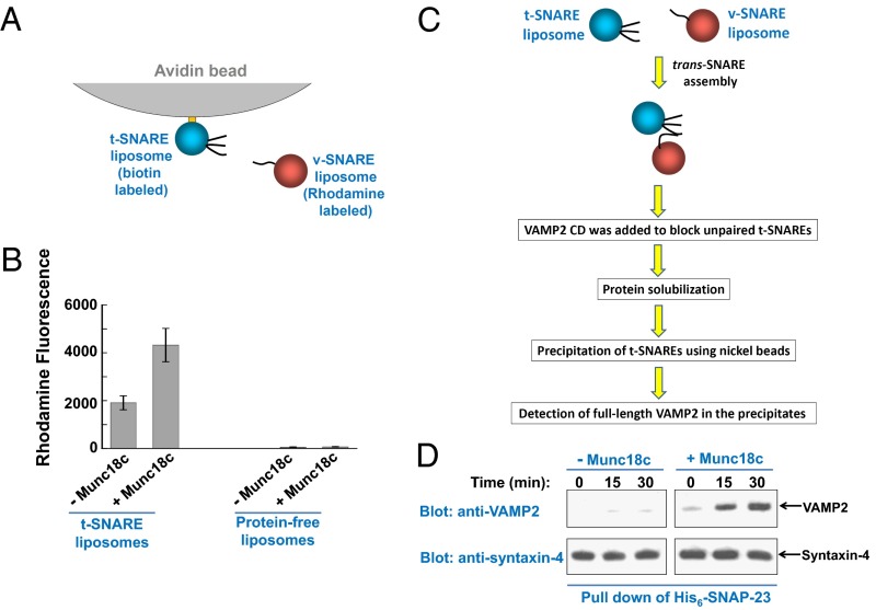
Fig. 3.
Munc18c promotes trans-SNARE zippering at the postdocking stage of the fusion reaction. (A) Diagram of the liposome pull-down assay. (B) Measurements of the docking of t- and v-SNARE liposomes using the liposome pull-down assay. Biotin-labeled t-SNARE liposomes were anchored to avidin agarose beads and were used to pull down rhodamine-labeled v-SNARE liposomes. The binding reactions were performed at 4 °C for 1 h in the absence or presence of 5 μM Munc18c. Data are presented as rhodamine fluorescence intensity. In the negative control, t-SNARE liposomes were substituted with protein-free liposomes. Error bars indicate SD. (C) Diagram of the trans-SNARE formation assay. (D) Reconstituted t- and v-SNARE liposomes were incubated at 4 °C for indicated periods in the presence or absence of 5 μM Munc18c before a 10-fold excess amount of inhibitory VAMP2 CD was added to block unpaired t-SNAREs. The liposomes were subsequently solubilized, and the t-SNAREs were precipitated using nickel Sepharose beads. The presence of full-length VAMP2 in the precipitates was probed by Western blotting, which was used as an indicator for trans-SNARE assembly between liposomes.

