
Fig. 1.
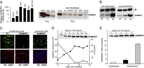
Expression of SAMHD1 in cultured human cells. (A) Relative levels of SAMHD1 mRNA and protein in different cell lines. The relative amount of mRNA was measured by RT real-time PCR in transformed cell lines (THP1, HEK293, and Jurkat) and in nontransformed lung and skin fibroblasts. SAMHD1 protein was detected by immunoblotting in extracts from transformed cells, lung and wild-type skin fibroblasts, and skin fibroblasts mutated for p53R2 or the mitochondrial thymidine (TK2) or deoxyguanosine (dGK) kinases. (B) Immunoblots of SAMHD1 from proliferating (P), confluent (C), and quiescent (Q) WT skin and lung fibroblasts. Asterisk marks an unspecific band (loading control). (C) Immunofluorescence shows nuclear localization of SAMHD1 (green) and cytosolic localization of the R1 (green) and R2 (red) subunits of RNR in lung fibroblasts. (D) Inverse relation between frequency of S-phase cells (filled circle) and abundance of SAMHD1 protein (open square) in proliferating cultures of lung fibroblasts. (E) Content of SAMHD1 in quiescent lung (black) and skin (gray) fibroblasts relative to the amount at confluency. The immunoblot shows the increasing SAMHD1 signal in lung fibroblasts.