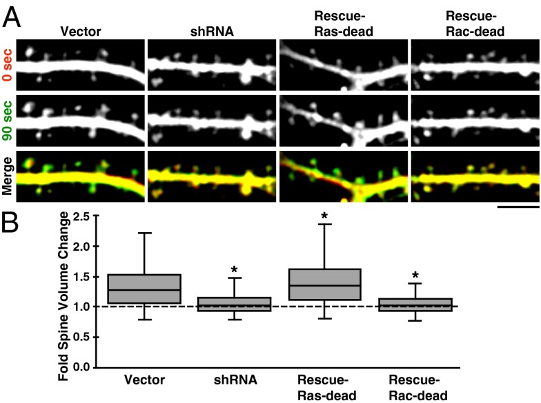
Fig. 5.
RasGRF2’s Rac-GEF activity is necessary for spine enlargement. (A) Stretches of dendrite and spines are shown from the indicated conditions demonstrating the degree of NMDA-induced spine enlargement. Images were assigned the same color as their time-label [i.e., before or 90 s after NMDA stimulation, when spine enlargement is maximal (Fig. 1B)] and merged in the bottom row; green-shifted spines are larger. Contrast enhancement as used for these images was not used for data analysis. (Scale bar, 5 µm.) (B) Box-and-whisker plot of spine volume changes between the 0- and 90-s timepoints (n > 500 spines pooled from n = 9 neurons per condition); see main text for details. Spine volume changes were diminished in the RasGRF2 knockdown and Rac-dead rescue conditions relative to vector. Ras-dead rescued spines enlarged slightly more than those in the vector condition, although the difference is significant because of the large number of spines analyzed. Whiskers represent data falling within 1.5 × IQR. Asterisks denote a significant difference from vector condition with P < 0.01.

