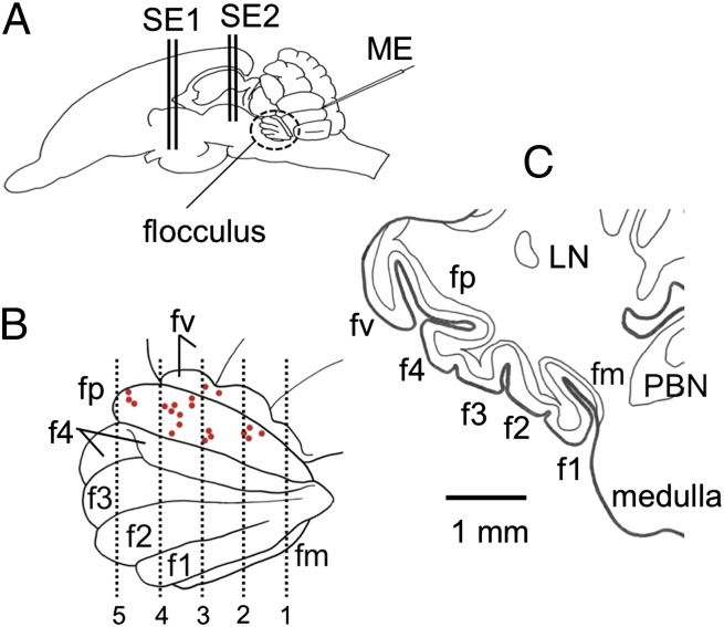
Fig. 1.
Folium-p of rabbit flocculus. (A) Left side view of rabbit brain. ME, glass microelectrode; SE1, stimulating needle electrode inserted in the hypothalamus; SE2, another electrode in PAG. (B) Enlarged left side view of the flocculus showing its folial divisions. fp is exposed by retracting parts of the neighboring f4 and fv. Red spots, positions of the last microelectrode tracks in 21 experiments. Vertical lines 1–5 are drawn at 400, 800, 1,200, 1,600, and 2,000 μm from the caudal pole of fp. (C) Coronal section of the left flocculus at the level 4 in B. LN, lateral nucleus.

