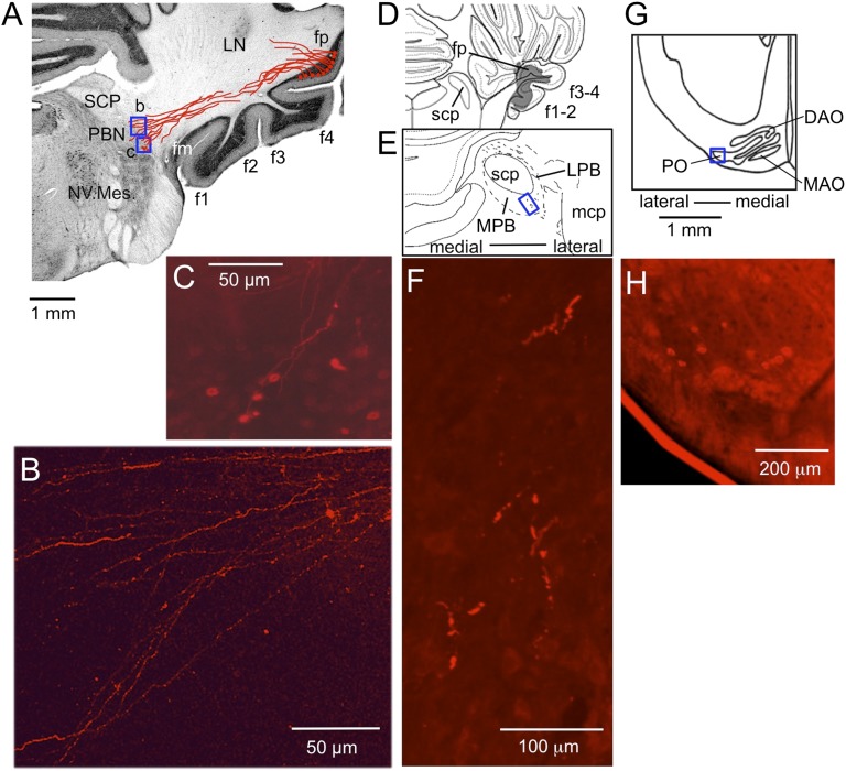
Fig. 6.
DiI- and BDA-labeled outputs and inputs of fp. (A) Coronal sections of cerebellum and medulla through right fp, into which DiI was injected. DiI-containing fibers were traced through three consecutive sections (100 μm thick) and superposed as drawn in red. LN, lateral cerebellar nucleus; NV. Mes., mesencephalic trigeminal nucleus; SCP, superior cerebellar peduncle. (B and C) Photomicrographs for two small areas (b and c) enclosed in A showing varicous pattern of the labeled axons (B) and retrogradely labeled neurons (C), respectively. (D) A section of the brainstem of another rabbit, relatively rostral corresponding to the level 5 in Fig. 4. BDA covered fp but extended also to the other floccular folia. (E) A 45-μm-thick section cut at 810 μm caudal to D, showing tissues around SCP in a larger scale. Wavy short lines indicate BDA-labeled axons. LPB, lateral PBN; MPB, medial PBN; mcp, middle cerebellar peduncle. (F) A photomicrograph of the part enclosed by a rectangle in E showing axon terminal-like structure labeled by BDA. (G) A section of the ventrolateral medulla at the caudal level of the principal olive (PO) of a third rabbit. Only the side contralateral to the fp injected with DiI is shown. (H) A photomicrograph for the area enclosed by a square in (G) showing retrogradely labeled IO neurons.

