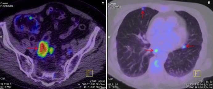
Figure 3.
(A) Positron emission tomography (PET) scan demonstrating circumferentially increased uptake in the wall of the rectosigmoid junction and adjacent lymph node, consistent with a primary colorectal carcinoma. (B) PET scan showing multiple subcentimeter nodules with increased uptake across both lung fields suggestive of metastatic disease.

