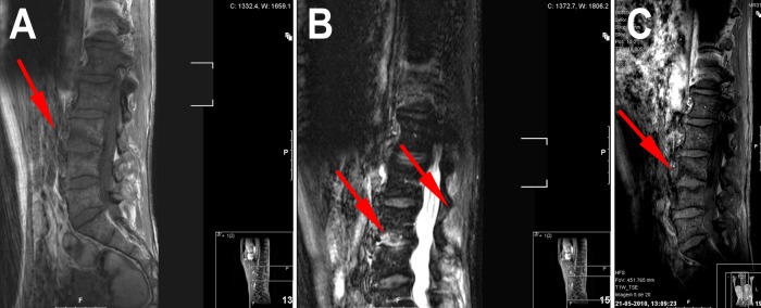
Figure 2.

Sagital MRI: T1 (A) shows at L3–L4 level a hypointense signal in the vertebral marrow adjacent to the inferior L3 endplate, with loss of endplate definition on the superior side of the disc. Axial stir (B) shows hyperintense signal in the endplate anal disc (anterior two thirds). Postgadolinium axial T1 (C) shows enhancing signal in the endplate.
