Abstract
A 33-year-old man presented with lymphoedema and obstructive nephropathy and was first diagnosed as retroperitoneal fibrosis (RF) with consistent clinical picture and radiographic findings. Further CT-guided biopsy was performed and non-Hodgkin lymphoma was diagnosed based on pathological results. RF is usually diagnosed through clinical presentation and imaging studies. However, our case proved that biopsy should be considered to exclude malignancy, even with typical presentations of RF. Follow-up after six courses of R-CHOP (rituximab, cyclophosphamide, vindesine, epirubicin and prednisone) regimen revealed complete resolution of symptoms.
Background
Retroperitoneal fibrosis (RF) is a rare inflammatory process, which involves predominantly middle-aged men aged between 40 and 60 years. Most cases of RF are idiopathic, commonly concurrent with other systemic autoimmune features. Recent study revealed that it may belong to the family of IgG4-related disease.1 Secondary RF is due to drugs, malignancy, infections or postradiation therapy. Although diagnosis of RF is usually based on clinical manifestation and imaging presentation, further investigation based on pathology and immunohistochemistry is sometimes needed to exclude other causes, especially malignancy. We hereby report a case of non-Hodgkin lymphoma in a patient presented with typical symptoms and signs of RF.
Case presentation
A 33-year-old man noticed a swollen lymph node in his left inguinal region in May 2007. It slowly enlarged to the size of a peanut within 1 year. In 2008, he underwent lymph node biopsy, which revealed only lymphatic reactive proliferation. However, a swelling in his left inguinal region spread gradually to the knee. It was aggravated by standing upright and partially relieved by lying supine. In 2010, the swelling spread to his left foot and his leg started to be painful. A lymphatic scan revealed lymphatic obstruction of the left thigh, and he was treated with troxerutin for 4 months, which showed no effect. In December 2011, the patient started to report of flank pain, oliguria and dark urine. He was diagnosed with RF in a local hospital after an abdominal CT scan was made. His medical history is not significant. He has been smoking 0.5 pack/day of cigarettes for 18 years.
Investigations
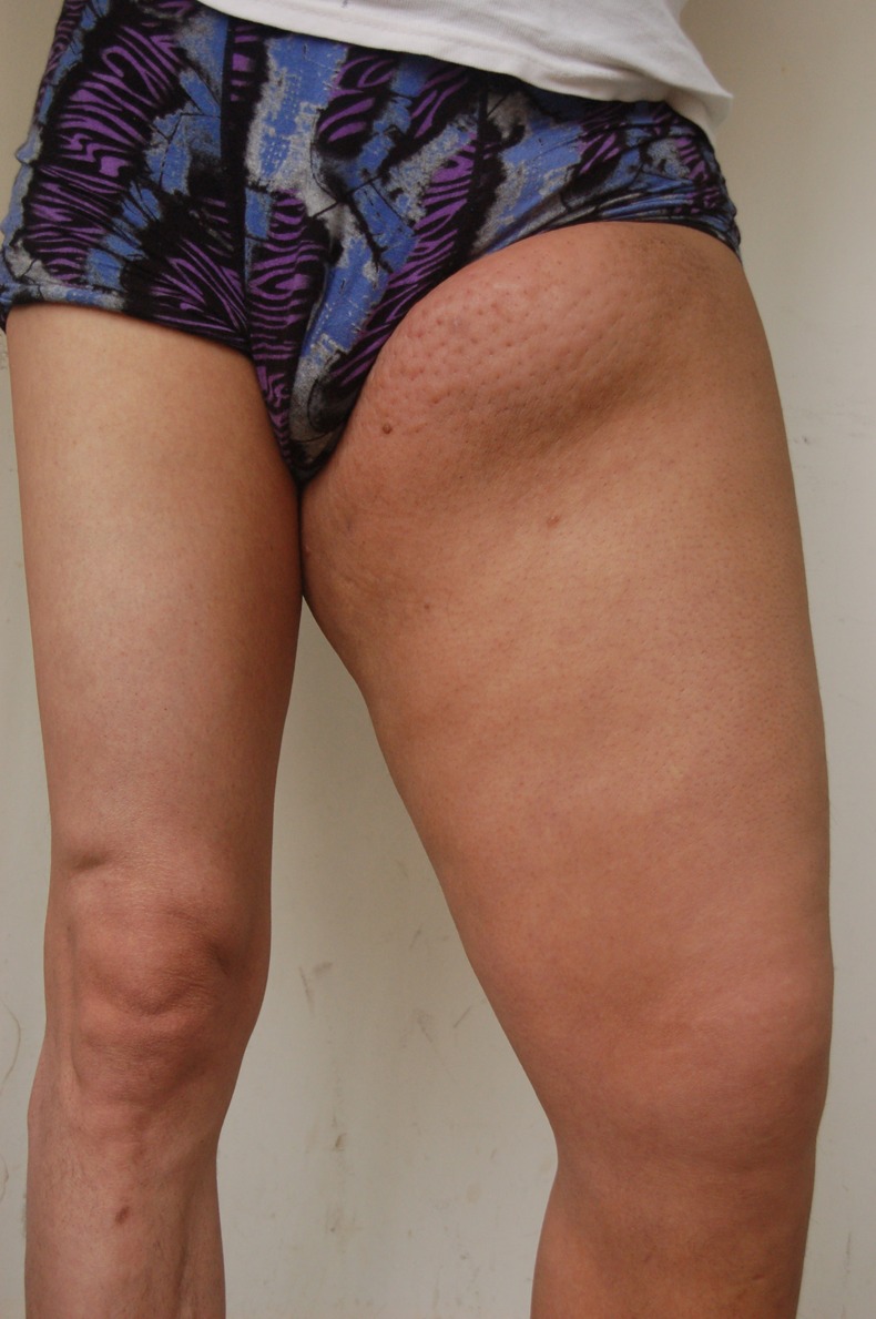
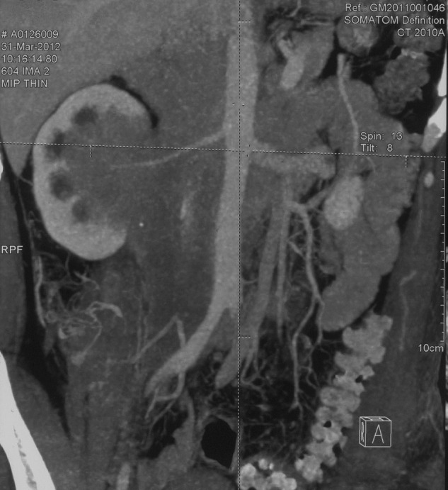
On admission, physical examination was significant for varicose veins on the upper abdomen, tenderness of middle upper quarter of abdomen, palpation of liver 2 cm below costal margin, shifting dullness and non-pitting oedema of the left lower limb (figure 1) with normal skin temperature. Complete blood count was normal and chemistry panel showed a urea of 5.58 mmol/L and serum creatine of 75 μmol/L. Twenty-four hour urine protein was 1.74 g. Autoimmune panels including antinuclear antibody, antineutrophil cytoplasmic antibody, antiextractable nuclear antigen antibody, anticardiolipin antibody, rheumatic factor were all negative. IgG and its subclass levels were within normal range. Vascular ultrasound revealed segmental occlusion of inferior vena cava, and hypoechogenic lesion around abdominal aorta, bilateral sacral arteries and right renal artery. Inguinal ultrasound found bilateral inguinal lymphadenopathy and a hypoechoic mass in the left inguinal region. Abdominal CT scan with contrast revealed a retroperitoneal soft-tissue mass, which invaded the right kidney and surrounded the abdominal aorta, inferior vena cava, superior mesenteric artery and bilateral renal vessels (figure 2). Left renal pelvic and bilateral psoas major muscles were also involved. CT urography revealed hydronephrosis, with occlusion of right renal pelvis and right ureter. A renal scan showed the glomerular filtration rate to be 9.46 (left) and 65.39 mL/min (right). CT-guided needle biopsy was performed. The results revealed fibrous and striated muscle tissue, infiltrated by lymphocytes. Immunohistochemistry analysis revealed CD20 (++), CD68 (+), CD138 (−) and IgG4 (−). Gene rearrangement revealed monoclonal activity of B-cell line. The patient was diagnosed as having non-Hodgkin lymphoma of B-cell type.
Figure 1.

Severe oedema of the left lower limb of the patient on admission.
Figure 2.

Coronal abdominal CT scan showing a retroperitoneal soft-tissue mass, encasing the abdominal aorta and invading the right kidney.
Differential diagnosis
Differential diagnosis of RF includes retroperitoneal malignancy, granulomatous infections and more rarely, retroperitoneal fibromatosis and inflammatory pseudomotor.
Histological and immunological investigations are sometimes needed to differentiate retroperitoneal fibrosis, and to suggest the presence of an underlying condition of secondary cases.
Treatment
On 2 May, a chemistry panel showed urea to be 10.05 mmol/L and serum creatine of 204 μmol/L. A double J stent was inserted the next day. His serum creatine decreased to 131 µmol/L and urea decreased to 6.27 mmol/L. Then chemotherapy started with R-CHOP regimen including rituximab, cyclophosphamide, vindesine, epirubicin and prednisone. His swelling of left thigh significantly improved.
Outcome and follow-up
The patient was last followed up in October 2012, after six courses of R-CHOP regimen. The double J stent was removed, and his symptoms resolved completely with normal renal function.
Discussion
RF is a rare inflammatory disease characterised by sclerosing retroperitoneal mass and obstructive clinical features. In most cases (80–100%), ureters are the uniformly involved retroperitoneal organs, causing hydroureter and hydronephropathy, even obstructive renal failure. The inflammatory mass can also compress arteries, veins and lymphatic vessels, resulting in leg oedema, deep vein thrombosis, hydrocele and claudication. Other symptoms also include non-specific constitutional symptoms such as fatigue, anorexia and weight loss.2 The diagnosis is largely dependent on characteristic imaging results, such as the homogeneous plaque surrounding the lower abdominal aorta on CT scan.3
About 70% of cases of RF are idiopathic, and often associated with other autoimmune conditions. Recently, IgG4-related disease has been found to be closely linked with RF. More than half of the cases of idiopathic RF present with significantly higher serum IgG4 level, along with IgG4-positive plasma cells in biopsy analysis. IgG4-related RF does not differ from IgG4-unrelated cases in clinical presentation or treatment outcome, except that most IgG4-related patients are men, while most IgG4-unrelated patients are women.4 Pathology of IgG4-related disease shows a striking similarity of mixed lymphoplasmacytic infiltration of B and T cells, and rich in IgG4-positive plasma cells.5 Other manifestations of IgG4-related disease may also be present, including a variety of fibrosclerosing disease. Commonly involved organs include thyroid, pancreas, biliary tree, mediastinum, pericardium and retroperitoneum.1
Our patient is a middle-aged man with distinct features of lymphoedema and obstructive nephropathy. Clinical features as well as CT results were consistent with RF. However, his serum IgG4 level was normal and further biopsy analysis confirmed the diagnosis of non-Hodgkin lymphoma. Non-Hodgkin lymphomas are always important entities to consider in differential diagnosis with RF. Their similarity in clinical and radiological presentation has posed a diagnostic challenge. Both of them have effective but very distinctive treatments, and lead to different prognosis. For idiopathic RF, long-term daily corticosteroid use is the primary treatment and the prognosis is usually good. Reported case series revealed a successful rate of 50–100% with prednisone monotherapy of 6 months to 2 years.6 On the other hand, lymphomas require specific chemotherapy protocols and have varying prognosis based on the prognostic index.
The subtle difference may be revealed via imaging tool. CT scan may find that idiopathic RF tends to encase the aorta and ureters, while retroperitoneal malignancy usually displace the aorta anteriorly and the ureters laterally.2 In our case, the CT imaging result revealed that the retroperitoneal mass in our patient invaded his right kidney (figure 2) and psoas major muscles in a more aggressive way than in idiopathic RF. These clues suggested the possibility of malignancy and did guide us to search for other causes of the retroperitoneal mass. Under MRI investigation, lymphoma more likely involves suprarenal and peri-renal location and presents with significantly greater size of the mass and lower value of the apparent diffusion coefficient7 However, these radiographic findings could only offer clues for the expert in this area, and for others these findings may be equivocal. Immunophenotyping of biopsied specimen may be critical to establish the definitive diagnosis.8 In our case, monoclonal populations of B cells are consistent with lymphoma.
To our knowledge, only few cases of lymphoma presenting as RF was reported in literature.9 10 Though imaging tools may help, biopsy is still necessary to differentiate from possible malignancy, especially in those who have little systemic autoimmune presentation. Even though the diagnosis of lymphoma was missed at the beginning; when RF responds poorly to steroid therapy, malignancy should also be considered and ruled out by biopsy.
Learning points.
Retroperitoneal fibrosis is identified by distinctive symptoms and radiological findings, usually involving obstructive features, most commonly ureteral obstruction. Lymphatic and blood vessel obstruction is also commonly seen.
Most retroperitoneal fibrosis cases present with other systemic autoimmune presentation. Many of them have elevated serum IgG4 concentration, along with IgG4-positive plasma cell infiltration in biopsy.
Differential between idiopathic retroperitoneal fibrosis and lymphoma is crucial since it alters the subsequent management and leads to very different prognosis.
Though imaging stools may help, biopsy is still necessary to differentiate possible malignancy, especially in those who have little systemic autoimmune presentation.
Footnotes
Contributors: NW and YJ collected the clinical information, treated the patient and wrote the manuscript.
Competing interests: None.
Patient consent: Obtained.
Provenance and peer review: Not commissioned; externally peer reviewed.
References
- 1.Vaglio A, Salvarani C, Buzio C. Retroperitoneal fibrosis. Lancet 2006;2013:241–51 [DOI] [PubMed] [Google Scholar]
- 2.Vivas I, Nicolás AI, Velázquez P, et al. Retroperitoneal fibrosis: typical and atypical manifestations. Br J Radiol 2000;2013:214–22 [DOI] [PubMed] [Google Scholar]
- 3.Zen Y, Onodera M, Inoue D, et al. Retroperitoneal fibrosis: a clinicopathologic study with respect to immunoglobulin G4. Am J Surg Pathol 2009;2013:1833–9 [DOI] [PubMed] [Google Scholar]
- 4.Clevenger JA, Wang M, Maclennan GT, et al. Evidence for clonal fibroblast proliferation and autoimmune process in idiopathic retroperitoneal fibrosis. Hum Pathol 2012;2013:1875–80 [DOI] [PubMed] [Google Scholar]
- 5.Stone JH, Zen Y, Deshpande V. IgG4 related disease. N Engl J Med 2012;2013:539–51 [DOI] [PubMed] [Google Scholar]
- 6.Rosenkrantz AB, Spieler B, Seuss CR, et al. Utility of MRI features for differentiation of retroperitoneal fibrosis and lymphoma. AJR Am J Roentgenol 2012;2013:118–26 [DOI] [PubMed] [Google Scholar]
- 7.Dash RC, Liu K, Sheafor DH, et al. Fine-needle aspiration findings in idiopathic retroperitoneal fibrosis. Diagn Cytopathol 1999;2013:22–6 [DOI] [PubMed] [Google Scholar]
- 8.van Bommel EF, Siemes C, Hak LE, et al. Long-term renal and patient outcome in idiopathic retroperitoneal fibrosis treated with prednisone. Am J Kidney Dis 2007;2013:615–25 [DOI] [PubMed] [Google Scholar]
- 9.Hammer ST, Jentzen JM, Lim MS. Anaplastic lymphoma kinase–positive anaplastic large cell lymphoma presenting as retroperitoneal fibrosis. Hum Pathol 2011;2013:1810–12 [DOI] [PubMed] [Google Scholar]
- 10.Chim CS, Liang R, Chan AC. Slcerosing malignant lymphoma mimicking idiopathic retroperitoneal fibrosis: importance of clonality study. Am J Med 2001;2013:240–1 [DOI] [PubMed] [Google Scholar]


