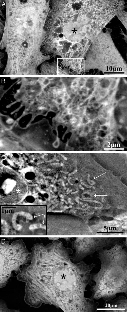
Fig. 2.
Imaging of stained cells. Asterisks denote nuclei, and thin arrows denote mitochondria. (A) HeLa cells grown on the membrane in normal growth medium, then fixed with paraformaldehyde, and stained with uranyl acetate (imaged at 12 kV). (B) Magnification of the marked rectangle shown in A. (C) CHO cell fixed with glutaraldehyde and paraformaldehyde, stained with uranyl acetate, and maintained in water (imaged at 30 kV). The thick arrow denotes a mitochondrion that surrounds a lipid droplet. (Inset) Higher magnification showing mitochondria (thin arrows). (D) Actin fibers in stained CHO cells. Treatment and imaging are as described for A.

