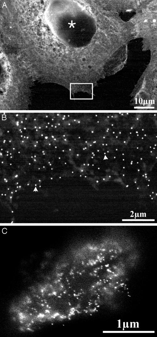
Fig. 3.
Imaging of nanoparticle gold markers on cells. The arrowheads denote gold nanoparticles. (A) Epidermal growth factor receptors immunolabeled with 40-nm gold nanoparticles on A431 cells and counterstained with uranyl acetate (imaged at 30 kV). (B) Magnification of the marked rectangle shown in E. (C) Putative gastrin receptors on H. pylori bacterium, after incubation with complexed biotinylated gastrin on streptavidin-coated 20-nm gold particles, followed by glutaraldehyde and sedimentation onto the poly-(l-lysine)-coated membrane (imaged at 20 kV).

