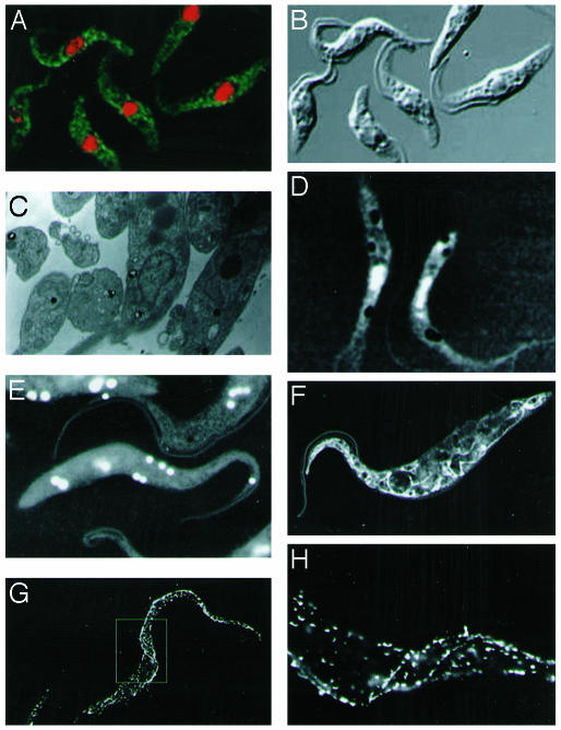
Fig. 4.
Multimodal imaging of T. brucei. The typical length of a cell is 20 μm in length and 3 μm in width. (A) Confocal fluorescence microscopy. Anti-EP procyclin antibodies (Cedarline) were used, and the binding was detected with anti-mouse linked to fluorescein isothiocyanate (green). Staining of the nucleus and kinetoplast was with propidium iodide (red). (B) Optical (differential interference contrast) imaging. (C) TEM sections of a cell, followed by uranyl acetate staining, revealing internal organelles. (D) Wet-SEM imaging of whole, hydrated, unstained cells. (E) As described for D using osmium tetroxide staining. (F) As described for D using uranyl acetate staining. (G) Nanoparticle gold markers linked to anti-mouse antibodies, showing the location of the surface EP procyclin protein and enabling at the same time a three-dimensional view. (H) Higher magnification of the midsection of the cell shown in G, elucidating an area of three-dimensional helicity in the cell.

