Fig. 5.
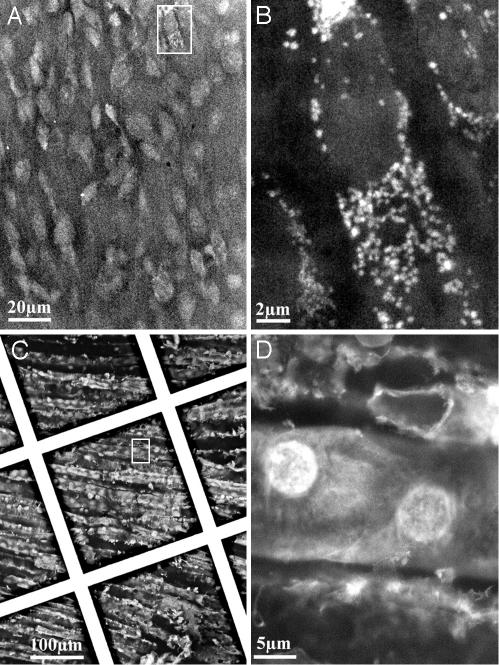
Imaging of tissues. (A) Mouse heart, untreated, imaged directly in the sample holder at 30 kV. (B) Higher magnification of the rectangle shown in A. (C) Rat kidney was dissected, cut, and fixed in formalin for 24 h. The tissue then was stained with 0.1% uranyl acetate for 10 min. C and D show epithelial cells within the inner surface of the tubules of the medullary rays. The grid supporting the membrane is visible in this picture. (D) Magnification of the box shown in C.

