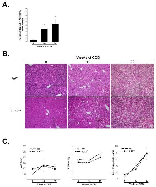
Figure 2. Increased IL12 production does not influence the progression of hepatosteatosis following long-term CDD feeding.
A. C57BL/6 mice were fed CDD for 0, 10 or 20 weeks. Hepatic IL-12 expression was determined by quantitative real time PCR of whole liver tissue while serum IL-12 was measured by ELISA. Values are means ± SEM,. B. H&E stained liver histology after 0, 10 and 20 weeks of CDD in C57BL/6 (Wt) and IL-12−/− mice. Representative photomicrographs are presented at 100x magnification with 400x inserts (bottom right of each image). C. Serum alanine aminotransferase, liver weight to body weight ratio (LW/BW) and hepatic triglycerides were measured following either 0, 10 or 20 weeks of CDD feeding. No significant differences were observed between Wt and IL-12−/− mice in any of these parameters. Values are means ± SEM, *p<0.05, n=4-8 animals per group.

