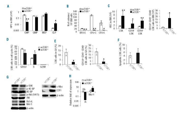
Figure 2.
Deletion of mTOR affects HSC and HPC homeostasis. (A) The numbers of common myeloid progenitors (CMPs) (Lin−Sca1−c-Kit+CD34+CD16/CD32mid), granulocyte-macrophage progenitors (GMPs) (Lin−Sca1−c-Kit+CD34+ CD16/CD32hi), megakaryocyte-erythroid progenitors (MEPs) (Lin−Sca1−c-Kit+CD34−CD16/CD32lo), and common lymphoid progenitors (CLP) (Lin−IL7R+Sca1medc-Kitmed-hi) in bone marrow (BM) were analyzed by FACS. (B) The colony-forming activities of CFU-E, CFU-C and BFU-E were examined using the total BM cells. (C) The numbers ofLSK (Lin− scal+c-kit+), CD34−LSK, CD34+LSK, and CD150+CD41−CD48−LSK cells in BM were analyzed by FACS. (D) Cell cycle profile of LSK cells. LSK cells were labeled with BrdU in vivo, followed by BrdU and 7-AAD staining and FACS analysis (G0/G1:BrdU−7-AADlo; S:BrdU+; G2/M:BrdU−7-AADhi). (E) The percentages of LSK progenitor cells and CD150+CD41-CD48-LSK stem cells in G0 phase (pyronin Y-Hoechst 33342lo) of cell cycle were determined. (F) The percentage of apoptotic LSK cells was analyzed by Annexin V staining. (G) Western blot analysis of the signaling activities in Lin- cells. (H) Quantitative RT-PCR analysis of LSK cells for the expression of indicated genes. n=3–5 in each test group. *P<0.05; **P<0.01. Error bars represent mean ± s.d.

