Figure 3.
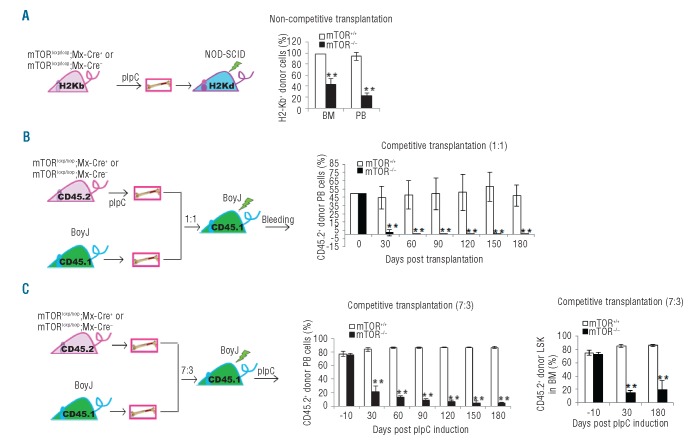
mTOR deficiency causes an impaired repopulating potential of HSCs. (A) Left: schematics of donor transplantation into NOD-SCID mice. Right: bone marrow (BM) cells from pIpC-treated donor mice were transplanted into sub-lethally irradiated NOD/SCID mice. The donor-derived cells in BM and peripheral blood (PB) of recipient mice were analyzed identified by H2-Kb+ staining. (B) Left hand side: schematics of competitive donor transplantation into syngeneic BoyJ mice. Right: BM cells from pIpC-treated donor mice (C57Bl/6) were mixed at 1:1 ratio with BM cells from BoyJ recipient mice. The cell mixtures were transplanted into lethally irradiated BoyJ rmice. The donor-derived cells were identified by CD45.2+ staining. (C) Left: schematics of competitive donor transplantation into syngeneic BoyJ mice. Middle and right: BM cells from donor mice were mixed at 7:3 ratio with BM cells from BoyJ recipient mice. The cell mixtures were transplanted into lethally irradiated BoyJ mice. The recipient mice were injected with pIpC 2 months post transplantation. The percentage of overall donor-derived cells in PB of the recipient mice were analyzed at various days post pIpC induction by CD45.2 staining (middle). The percentage of donor-derived LSK cells in BM of the recipient mice were determined at 30 days and 180 days post pIpC induction (right). N=3–5. **P<0.01. Error bars represent mean ± s.d.

