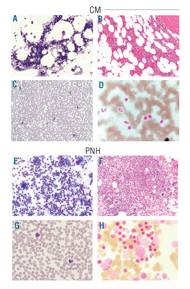
Figure 2.

Morphology of bone marrow and peripheral blood from SF3B1 mutated cases. (A) In the CM patient, bone marrow (BM) aspirate shows normal M:E ratio (Wright stain: ×20) and unremarkable morphology. (B) BM core biopsy shows slightly decreased cellularity for age (40–50%) (H&E stain: ×20) and (C) peripheral blood smear shows no remarkable features. (D) Prussian blue staining shows RS (6%) in the BM. (E) In the PNH patient, BM aspirate shows remarkable erythroid hyperplasia (M:E ratio of 0.3) with megaloblastoid changes and occasional erythroid precursors with irregular nuclear contours or double nuclei (Wright stain: ×20). (F) BM core biopsy shows 95% cellularity with erythroid hyperplasia (H&E stain: ×20). Megakaryocytes are present in adequate number. (G) Peripheral blood smear reveals mild normocytic anemia with moderate polychromasia and anisopoikilocytosis including ovalocytes and occasional fragmented red cells. (H) Prussian blue staining demonstrates slightly increased RS (17%) in the BM. CM: cutaneous mastocytosis; PNH: paroxysmal nocturnal hemoglobinuria; H&E: hematoxylin & eosin; M: myeloid; E: erythroid.
