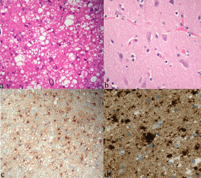
Figure 2.
Anatomopathological findings. (A) H&E stain showing diffuse typical spongioform changes in patient's cerebral cortex due to the presence of intracellular coalescent vacuoles (×400); (B) The aspect of the normal cortex is shown on the right for comparison (×400). (C) Immunohistochemistry showing marked gliosis (antiglial fibrillary acid protein antibody, ×200) and (D) plaque-like prion protein deposits. Test performed with monoclonal antiprion protein antibody POM1 at 1/200 dilution.

