
Figure 3.
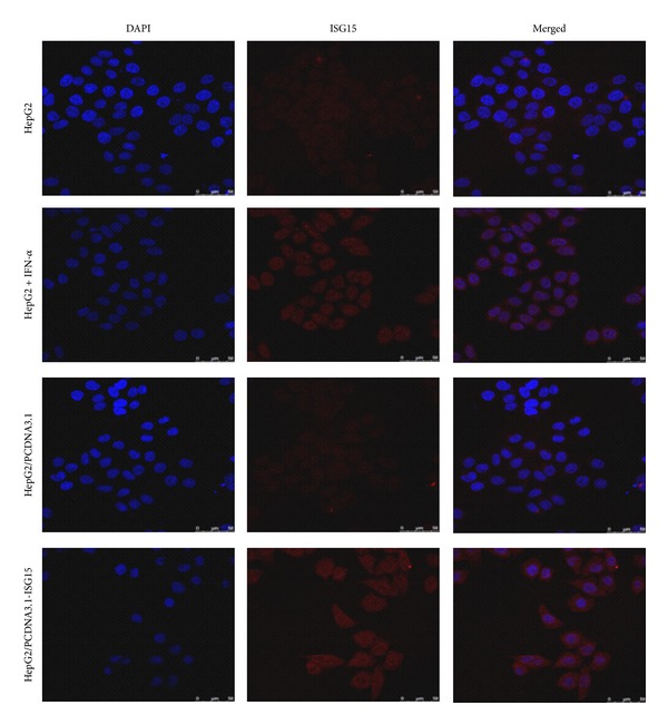
Subcellular localization of ISG15 in HepG2 cells. Free ISG15 and ISG15 modified proteins were detected by confocal-scanning laser microscopy at a magnification of 100x using anti-ISG15 antibody and Cy5-labeled secondary antibody. The Cy5 wavelength was detected by a confocal-scanning laser microscope and is shown in red, while nuclei were stained by DAPI and are shown in blue.