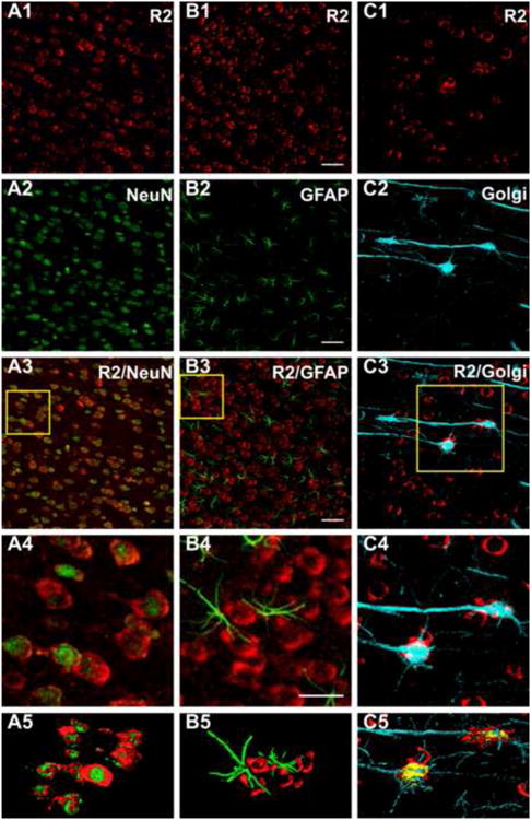
Fig. 5. 5αR2 co-localizes in pyramidal neurons but not in glial cells of the rat prefrontal cortex.

CLMS images showing strong 5αR2-immunolabeled neurons (R2) (red) (A1, B1, C1) and NeuN (A2-A5) and GFAP (B2-B5) (green) immunofluorescence in the prefrontal cortex (PFC). A3: merge of A1 and A2; B3: merge of B1 and B2; A4 and B4: higher power of the respective boxed area displaying the presence of 5αR2 immunoreactivity throughout the cytoplasm of neuronal cells (A4), but not in glial cells (B4). C2: Golgi-Cox impregnated pyramidal neurons. C3: merge of C1 and C2; C4: higher power of the boxed area displaying the co-localization of R2 with Golgi-Cox impregnated pyramidal neurons. A5, B5, C5 channels surface rendering for R2 and NeuN (A5), for R2 and GFAP (B5) and R2 immunofluorescence and Golgi-Cox (C5). Scale bars: 50 μm in A1-C3, 100 μm in A4, B4, C4. For interpretation of the references to color in this figure legend, the reader is referred to the web version of the article.
