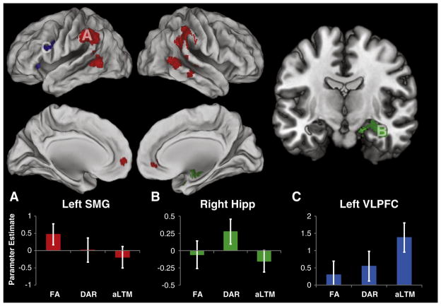
Fig. 3.
Neural correlates of access to visual short-term memory. Top: areas showing significantly different activation during the access of different putative states of visual short-term memory (p<0.05 FWE corrected). Red: areas selectively active for accessing the focus of attention (FA). Green: areas selectively active for accessing the direct access region (DAR). Blue: areas selectively active for accessing the activated portion of long-term memory (aLTM). Bottom: parameter estimates drawn from statistically independent regions-of-interest (ROI) for each contrast of interest (see Supplemental Methods for ROI details). A) Parameter estimates drawn from the left supramarginal gyrus (SMG) demonstrating selectivity for accessing the FA. B) Parameter estimates drawn from the right hippocampus (hipp) demonstrating selectivity for accessing the DAR. Section cut at y=−10. C) Parameter estimates drawn from the left ventrolateral prefrontal cortex (VLPFC) demonstrating selectivity for accessing the aLTM. For more detailed visualization of these activation patterns, see Supplemental Fig. 2.

