
Figure 4.
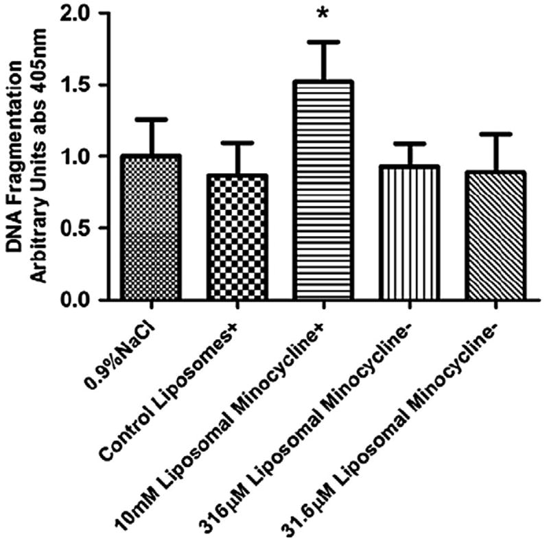
Level of fragmented DNA found in rat retinal lysates as detected with the Roche Cell Death Detection enzyme-linked immunosorbent assay. There was no increase in fragmented DNA detected when the animals were treated only with control nanoliposomes or nanoliposomal minocycline at concentrations of 316 μM or 31.6 μM minocycline as compared to animals treated with 0.9% NaCl irrigation fluid. The control and the 10 mM minocycline treatments were not exposed to CL-4B size-exclusion chromatography that removes surface-contaminated minocycline (+). In previous experiments (data not shown), this 10 mM free/nanoliposomal minocycline preparation was observed to induce apoptosis and here serves as a positive control. However, the equivalent preparation of liposomes without minocycline causes no such increase in DNA cleavage at this concentration and with this treatment regimen. 316 μM and 31.6 μM nanoliposomal minocycline preparations were purified via CL-4B size-exclusion chromatography (−). This step eliminates nonencapsulated minocycline and greatly reduces the concentration of minocycline in the preparation. In this experiment, increases in DNA cleavage were not detected in rat retinal lysates from rats treated with these preparations. Asterisk denotes statistical significance between the marked column and the retinal samples treated with 0.9% NaCl irrigation fluid as determined by a one-tailed nonparametric Mann-Whitney t-test (*α = 0.05).