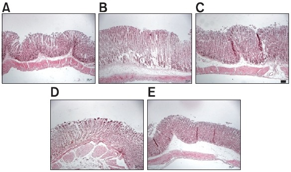
Fig. 3. Histological evaluation of the protective effects of WER and EER with hematoxylin and eosin (HE) stained longitudinal sections through gastric mucosa in an HCl/ethanol-induced gastric ulcer model. (A) Sham control group, (B) vehicle group, (C) positive control group treated with sucralfate, 100 mg/kg, (D, E) the stomach of test groups treated with WER and EER, 100 mg/kg, p.o., 1 hr before administration of 150 mM HCl/ethanol, respectively. A scale bar represents 150 μm.

