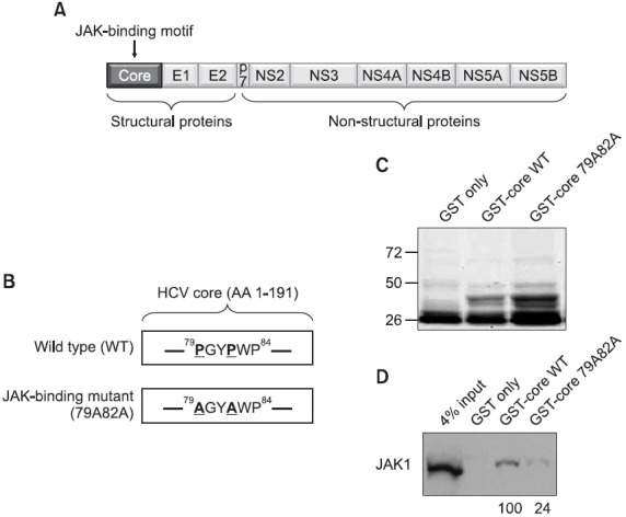
Fig. 1. (A) A schematic diagram of the HCV genomic map. The JAK-binding motif is found in the middle of the core protein region. (B) The amino acid sequence of the JAK-binding domain of the core protein. The critical two prolines in the JAK-binding motifs are highlighted with bold and underlines. The JAK-binding mutant core possesses the mutated two alanines in place of the two prolines in the JAK-binding motif. (C) 1 μg of purified GST, GST-core WT, and GST-core 79A82 proteins were separated by SDS-PAGE and stained with a brilliant coomassie blue. (D) Interaction of the HCV core protein with the JAK1 through the JAK-binding motif of the core protein. Either GST-core WT or GST-core 79A82A mutant proteins were mixed with liver carcinoma Huh7 cell lysates followed by Western blot analysis using an anti-JAK1 antibody. The numbers below the blot indicate the relative levels of recovered JAK1 proteins in each experiment.