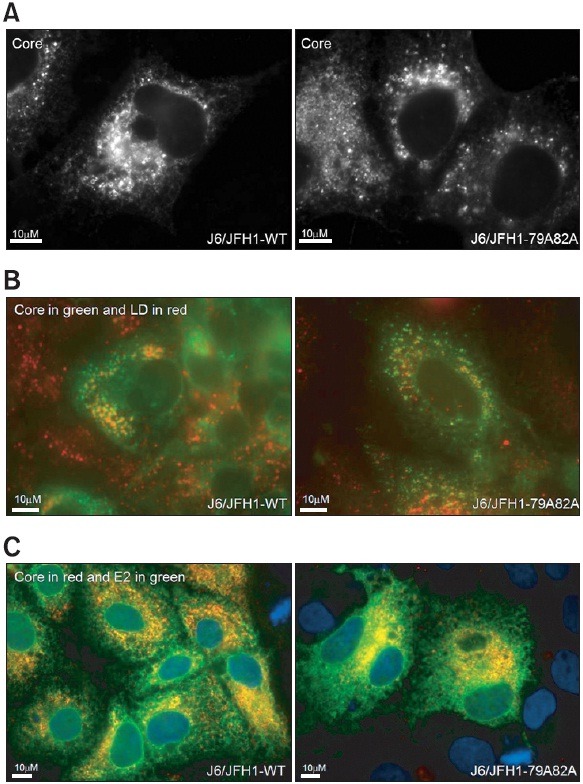
Fig. 6. (A) Immunofluorescence staining of core proteins in the cells transfected with either wild type J6/JFH1 or mutant J6/JFH1-79A82A RNAs at 3 days post-transfection. The viral RNAs-transfected cells were staining with a mouse anti-core antibody and an anti-mouse Alexa 488 antibody. (B) Immunofluorescence staining of both core proteins and lipid droplets in the cells transfected with either wild type J6/JFH1 or mutant J6/JFH1-79A82A RNAs at 3 days post-transfection. The RNAs-transfected cells were staining with a mouse anti-core antibody and an anti-mouse Alexa 488 antibody. Lipid droplets were stained with an oil red O dye. (C) Immunofluorescence staining of both core proteins and HCV envelope glycoprotein E2 in the cells transfected with either wild type J6/JFH1 or mutant J6/JFH1-79A82A RNAs at 3 days post-transfection. The RNAs-transfected cells were staining with a mouse anti-core antibody and an anti-mouse Alexa 594 antibody followed by a rabbit anti-E2 antibody and an anti-rabbit Alexa 488 antibody. Nucleus in each cell was stained with DAPI.