. 2013 Mar;21(2):107–113. doi: 10.4062/biomolther.2012.102

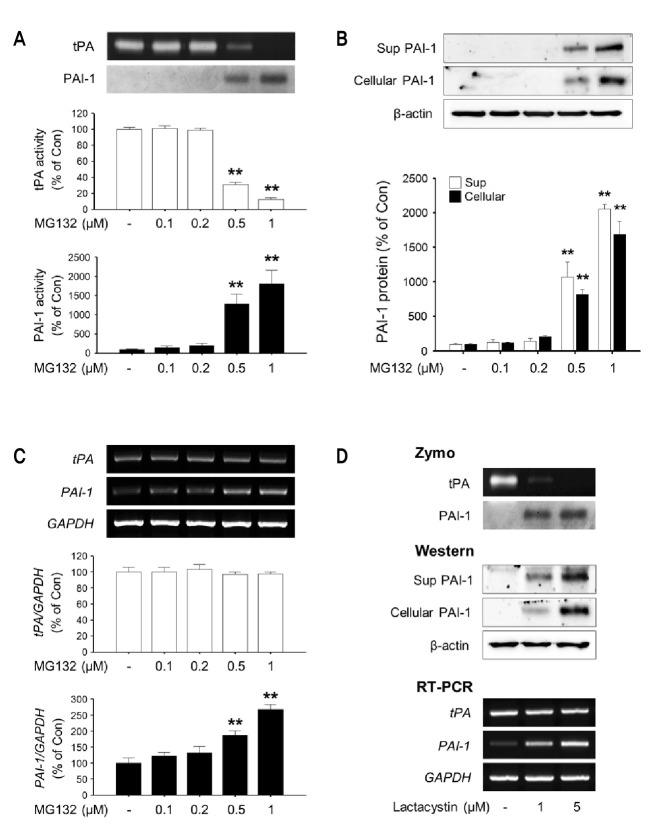
Fig. 1. The effects of MG132 and lactacystin on tPA/PAI-1 activity, protein and mRNA levels in rat primary astrocytes. Rat primary astrocytes were incubated in serum-free DMEM/F12 with increasing concentration of MG132 (0.1-1 μM) for 24 h. (A) The activity of tPA/PAI-1 in the culture supernatant was determined by casein zymographic procedure as described in Materials and Methods. (B) The level of PAI-1 protein secreted into culture spent media (Sup) and cell lysates (Cellular) were determined by Western blot using specific antibody against PAI-1. β-Actin levels were measured as a loading control. (C) Expression of total cellular tPA and PAI-1 mRNA were measured by RT-PCR. GAPDH levels were measured as an internal control. (D) Rat primary astrocytes were incubated in serum-free DMEM/F12 with Lactacystin (1, 5 μM) for 24 h. The activity of tPA/PAI-1 in the culture supernatant, the amount of PAI-1 protein secreted into culture spent media and cell lysates, expression of total cellular tPA and PAI-1 mRNA were determined as described. All graphs represent the mean ± S.E.M. of four independent experiments (**p<0.01 versus Control).

Fig. 1.