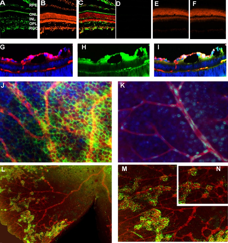
Figure 1.
Unfolded protein response is activated in the ERAI OIR retina. (A–F) The expression of the spliced Xbp1-GFP was detected in retinal cryosections in the RGCs, outer plexiform layer, outer nuclear layer, and inner nuclear layer in P17 ERAI OIR compared with naive ERAI. Nuclei were stained with propidium iodide (red), and the GFP protein (green) was detected by immunohistochemistry with antibody against GFP to avoid autofluorescence and increase our specificity of detection. (E, G, H, I) Formation of retinal blood vessels in the ERAI OIR mice. The retinal cryosections were stained overnight with lectin, a blood vessel marker (red). Detection of the GFP protein was observed in pericytes (green). Nuclei were stained with DAPI (blue). (J, K) The pericytes of the ERAI OIR blood vessel (J) experience the activation of the IRE signaling and the splicing of Xbp1 compared with pericytes in control ERAI retina (K). (L) Immunostaining of the ERAI IOR flat-mounted retina was performed using anti-lectin and anti-GFP antibodies; nuclei were stained with DAPI. (M) and (N) are high-resolution images of (L).

