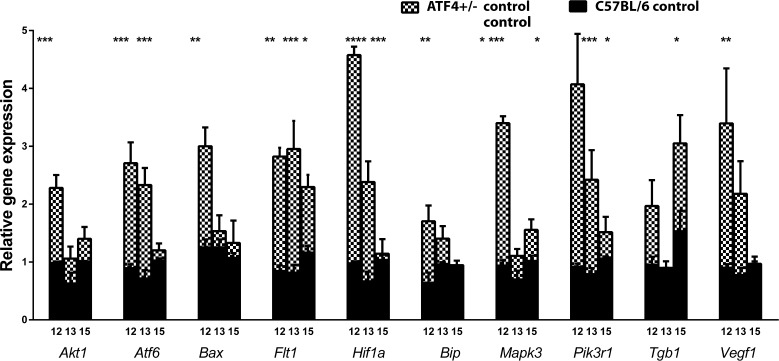
Figure 3.
Alteration of UPR-induced and vascularization-related gene expression during retinal development in ATF4+/− mice. The relative gene expression was articulated in RQs and was calculated in C57BL/6 (n = 4) and ATF4+/− (n = 4) naive animals at P12, P13, and P15 using C57BL/6 controls as a reference at each time point. Two-way ANOVA was used to calculate the difference between groups. At P12, we observed a 2.3-fold increase in Akt, a 3.0-fold increase in Atf6, a 2.4-fold increase in Bax, a 2.7-fold increase in Bip, a 3.6-fold increase in Mapk3, and a 4.6-fold increase in Hif1a gene expression. At P13, the expression of these genes was back to control levels with the exception of Hif1a and Atf6; their expression was still elevated by 3.2-fold and 3.6-fold, respectively. At P15, the expression of UPR-induced genes did not differ from that of controls. Vascularization-related gene expression was also altered in ATF4+/− retinas during retinal development. For example, Pik3r1 expression was increased by 4.4-fold and 3.1-fold at P12 and P13, respectively. By P15, its expression was back to control levels. While Vegfa and Tgfb1 expression was upregulated only at P12 and P15 by 3.7-fold and 2.0-fold, respectively, the expression of Flt1 was consistently elevated during the analyzed period and was 3.3-fold, 3.6-fold, and almost 2.0-fold higher in ATF4+/− mice at P12, P13, and P15, respectively. See also Supplementary Table S1 (*P < 0.05, **P < 0.01, ***P < 0.001, ****P < 0.0001).

