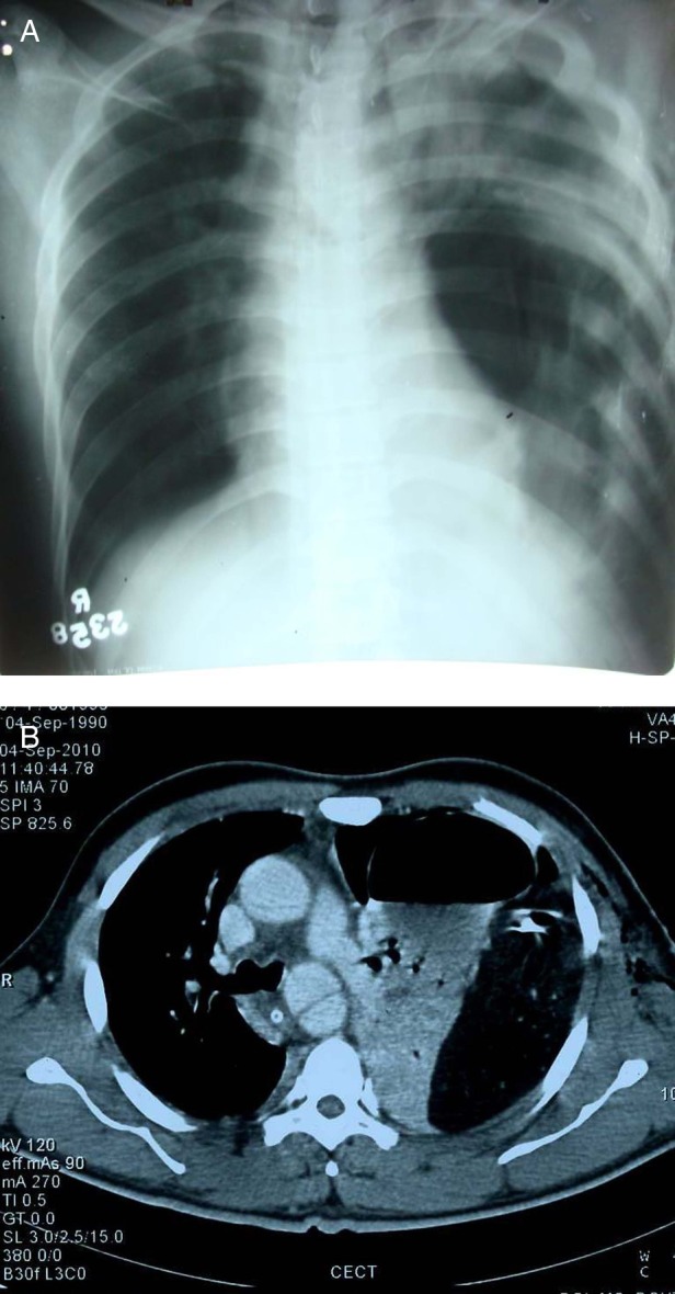
Figure 3.

(A) Radiograph of the chest posteroanterior view reveals loculated pneumothorax in the left hemithorax with mild mediastinal shift. Haziness is also seen on the left side which was misinterpreted as motion blur due to breathing. However, in retrospect it was possibly due to peristalsis. (B) Axial CT scan of the same patient at the level of aortic arch reveals herniation of stomach and mesentry into the left hemithorax and ‘dependent viscera’ sign.
